

Thinning bones in the upper spine may serve as an early, silent alarm system for accelerated brain aging and cognitive decline.
Getting older usually brings a few predictable creaks and groans, along with the occasional misplaced set of car keys. Global health trends from the National Institutes of Health show that bone loss is already a massive public health challenge, with osteoporosis affecting 18.3% of the world’s population and roughly 200 million women worldwide. In the United States alone, the Centers for Disease Control and Prevention reports that 12.6% of adults aged 50 and older have osteoporosis, while another 43.1% live with low bone mass.
Groundbreaking research published in the journal Radiology shows that skeletal fragility and mental sharpness are more closely linked than doctors previously realized. Led by Dr. Shadpour Demehri at Johns Hopkins University, the study tracked 715 older adults over five years and linked weaker upper-spine bones directly to faster white matter decay. “This comprehensive study using both imaging and clinical assessments sends a clear signal that bone density at baseline is associated with both functional and imaging measures of age-related brain degeneration,” Dr. Demehri explained.
Routine Chest Scans Unlock Hidden Brain Clues
Artificial intelligence can now repurpose routine chest scans to measure spinal strength and flag hidden neurological risks at no extra cost to patients.
Doctors order millions of noncontrast chest CT scans every year to check for lung nodules, heart calcium, or respiratory issues. Instead of ordering expensive new bone tests, the Johns Hopkins team used a validated deep-learning algorithm to automatically read bone mineral density from upper-spine vertebrae visible on those existing chest scans. That clever approach turns a standard heart or lung checkup into a two-for-one screening tool.
The investigation analyzed data from the Multi-Ethnic Study of Atherosclerosis between September 2016 and March 2024. Researchers followed 715 adults, 397 men and 318 women, with a median age of 69, who started the study completely free of dementia, stroke, or clinical heart disease. Over roughly five years, over 400 participants completed repeat brain MRIs, and more than 600 took serial cognitive tests to track mental sharpness.
“Chest CT is done for lung cancer screening, calcium scoring, nodule follow-up, and many other indications,” noted study co-author Dr. Sara Momtazmanesh. “These scans could provide an opportunistic measurement of bone density from the thoracic spine that is associated with a loss of cognitive function.” Dr. Demehri added that AI can synthesize immense amounts of imaging data across multiple organ systems, connecting age-related conditions that scientists traditionally studied in isolation.
Related: 10 Simple Habits to Recharge Your Mind and Body
Fraying Wires And Fading Sharpness
Patients with lower baseline spinal bone density experienced significantly faster deterioration in deep-brain wiring and overall thinking skills over the five-year tracking period.
To understand what happens inside an aging head, the research team examined two key MRI markers of brain wiring. First, they measured fractional anisotropy, which gauges how well white matter nerve tracts are organized. Second, they tracked white matter hyperintensities, which show up as bright spots on scans and signal small-vessel tissue damage.
Surprisingly, the total brain-wide drop in white matter organization did not hold up after strict statistical corrections. Instead, the damage hit specific, high-traffic hubs inside the brain. Lower bone density strongly predicted faster fraying of nerve fibers in the anterior limb of the internal capsule, a deep-brain cable linking movement and thinking centers.
At the same time, bright-spot tissue damage in the corpus callosum, the main bridge connecting the left and right hemispheres, increased by 12.8% each year. That structural wear and tear translated directly into lower mental test scores, even though isolated short-term digit-span memory stayed intact. Interestingly, participants with the weakest bones had higher education levels and healthier “good” HDL cholesterol (57.0mg/dL versus 53.0mg/dL), so statisticians had to adjust heavily for lifestyle, genetics, and heart risks to uncover the true biological trend.
Save this article
Why Diabetes Pours Fuel On The Fire
Having diabetes dramatically worsens the link between fragile spinal bones and small-vessel brain damage.
When researchers separated participants by blood sugar health, a striking pattern emerged. Among older adults with diabetes, lower spinal bone density went hand-in-hand with nearly 5% faster accumulation of white matter lesions. In people without diabetes, that link between thinning bones and bright-spot brain damage practically vanished.
The study authors suspect chronic inflammation and damage to the blood vessel lining, known clinically as endothelial dysfunction, explain why diabetes magnifies the threat. When high blood sugar batters tiny blood vessels, both bone tissue and deep-brain wiring starve for nutrients at the same time. That makes routine chest CT bone checks especially valuable for flagging high-risk diabetic patients early.
How Bones Whisper To The Brain
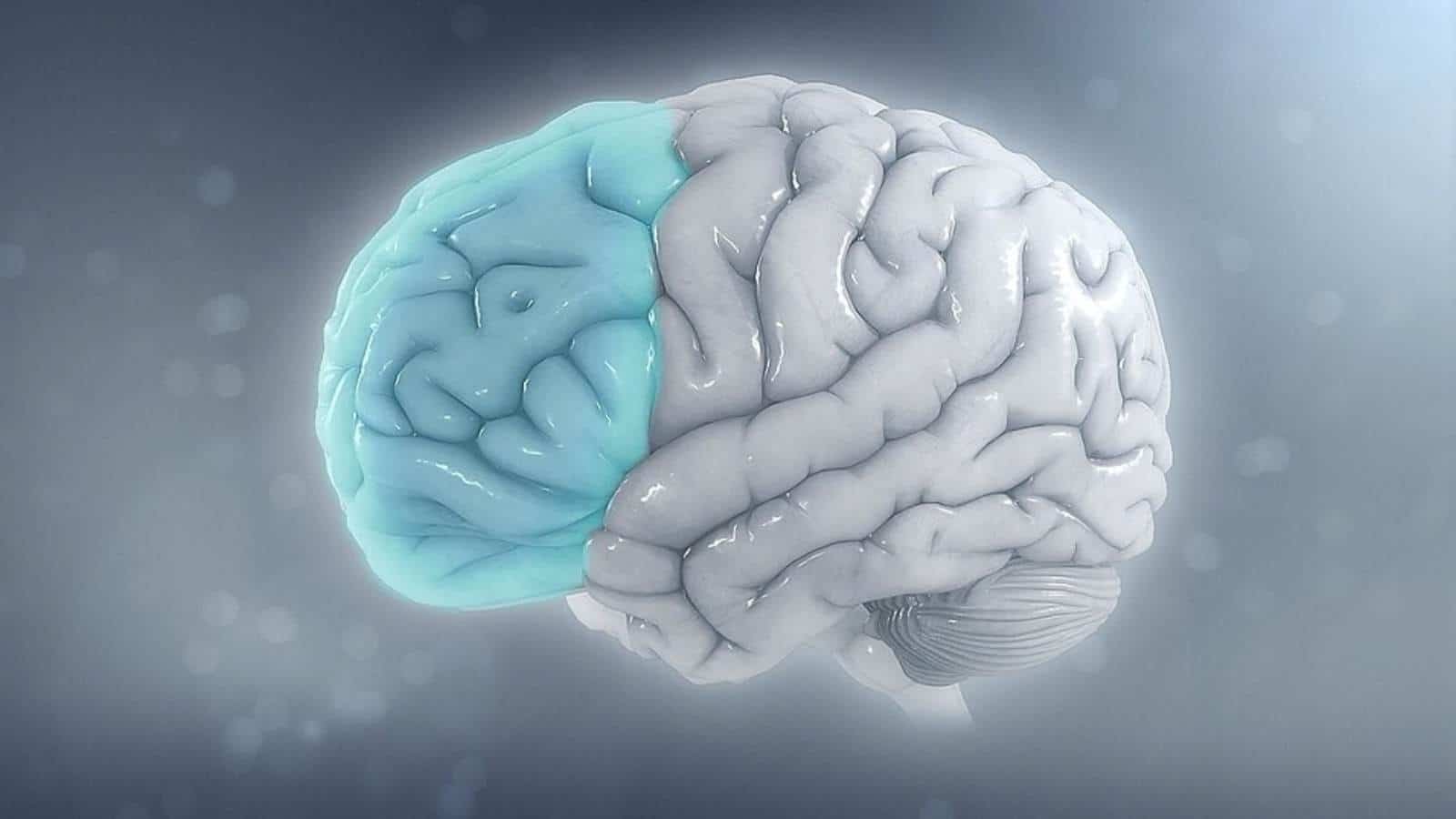
Weak bones don’t directly break the brain; instead, they reflect shared metabolic wear and tear and active chemical messaging between the skeleton and nervous system.
Scientists once treated the skeleton as lifeless scaffolding, but modern biology shows it is a lively endocrine organ. Bone-building cells release active proteins, such as osteocalcin, directly into the bloodstream. Research shows osteocalcin crosses into the brain, binds to specialized GPR158 receptors, regulates neurotransmitters, and even helps clear toxic amyloid plaques in animal models.
Aging bone marrow also undergoes biological shifts that mirror protein buildup and nerve cell breakdown in the brain. Still, Dr. Demehri stresses that the study is strictly observational and cannot prove that weak bones cause dementia. “This study is not about cause and effect, but rather the observation of a metabolic syndrome that may cause both bone and brain degeneration,” Dr. Demehri clarified.
Shared aging culprits, including insulin resistance, abnormal cholesterol, chronic inflammation, and menopausal hormone shifts, likely erode bone and brain tissue in parallel. Furthermore, the five-year window was too short to track full-blown dementia diagnoses, and researchers did not measure blood bone-turnover markers directly. The AI software also needs broader clinical validation across different CT scanner models before hospitals adopt it as a standard diagnostic tool.
The Skeletal-Cognitive Bottom Line
Protecting metabolic and skeletal health today may be one of the smartest ways to safeguard mental sharpness tomorrow.
This landmark Johns Hopkins study proves that the spine and brain age on a shared biological track. By applying AI to routine chest CT scans, doctors could soon spot early warning signs of cognitive decline years before memory slips become obvious.
While thinning bones don’t directly cause brain damage, they serve as a powerful dashboard light for whole-body metabolic health, especially for adults managing diabetes.
How might routine AI bone screenings during standard checkups change the way society approaches healthy aging, and what daily habits matter most for keeping both the spine and brain resilient?