

There is a strange kind of trust involved in going under general anesthesia. One moment you are answering a nurse’s questions beneath bright operating room lights. Then, seemingly seconds later, hours have passed, the surgery is finished, and you wake up with no memory of what happened in between. No sounds. No sensations. No dreams. Just a blank space.
For more than a century, the simplest explanation was that anesthesia was a form of deep sleep. That comparison was useful, but scientists now know it leaves out much of what is happening inside the brain.
New research suggests the anesthetized brain doesn’t simply switch off. Instead, it may move through a series of unusual states, with some areas still processing information while others become disconnected.
General anesthesia has been used in hundreds of millions of surgeries worldwide since the first successful public demonstration in 1846. Yet researchers are still working to understand exactly how it removes conscious awareness. Recent studies from Yale, a propofol trial published in Cell Reports Medicine, and a Baylor College of Medicine team recording individual neurons during surgery are revealing a much more complex picture.
Not Quite Sleep. Not Quite a Coma. Something Else Entirely.
The familiar shorthand was always a simplification. Research published in May 2026 by Yale is starting to show how much was being glossed over.
Janna Helfrich, a researcher at Yale School of Medicine, and her colleagues studied brain activity in propofol-sedated patients by comparing EEG recordings with patterns linked to deep sleep, REM sleep, coma, and wakefulness.
Instead of examining only the front of the head, as is common in many clinical settings, the researchers used 20 electrodes placed across the full skull to capture a broader view.
The results suggested that anesthesia does not create one single brain state. Different regions can show different patterns at the same time. Some activity looks similar to sleep, while other patterns resemble coma. Yet the anesthetized brain also appears to have characteristics that do not fit neatly into either category.
“The old dichotomy that it’s either sleep or coma is not true,” Helfrich said. “It’s actually both sleep and coma, and can be similar to both states at the same time, depending on where you look. And yet, there is also an element which is just anesthesia uniquely.”
After more than 150 years of use, scientists are still learning how the brain changes during the journey from awareness to unconsciousness.
Related: 8 Everyday Mistakes That Quietly Damage Brain Health
A Signal Hiding Inside the Brain Waves
A study of 31 people receiving propofol before surgery may have caught the brain-wave fingerprint of the moment consciousness disappears.
In a study published in Cell Reports Medicine, researchers used 128 scalp electrodes on each participant and tracked signals from nine brain regions involved in conscious experience. Before propofol took effect, activity between the parietal cortex and the thalamus showed strong coordination through alpha-band rhythms. As anesthesia deepened, that connection weakened.
Ti-Fei Yuan of Shanghai Jiao Tong University described the finding as opening “the possibility of a translatable biomarker,” meaning a measurable signal that could eventually help doctors adjust anesthetic doses more precisely. The goal would be to provide enough medication to prevent awareness while avoiding unnecessary depth.
The findings are not yet ready for routine medical use, but they show how closely researchers can now examine the transition into unconsciousness.
Save this article
What the Hippocampus Was Quietly Processing
The most unexpected result in recent months did not come from brain-wave measurements. It came from individual neurons and what they were doing while a patient was completely unconscious.
Researchers at Baylor College of Medicine used Neuropixels probes to record activity from individual neurons in the hippocampus of patients undergoing epilepsy surgery under general anesthesia. While the patients were fully sedated, researchers played unexpected tones and short spoken stories.
The hippocampus responded in ways that suggested surprisingly complex processing. Neural activity patterns could distinguish between nouns, verbs, and adjectives. The researchers also found signs that brain activity could anticipate upcoming words before they were spoken.
“The brain appears to anticipate what comes next in a story, even without conscious awareness,” said Sameer Sheth of Baylor. His colleague Benjamin Hayden added: “This kind of predictive coding is something we associate with being awake and attentive, yet it’s happening here in an unconscious state.”
The Brain’s Connections May Matter More Than Any Single Region
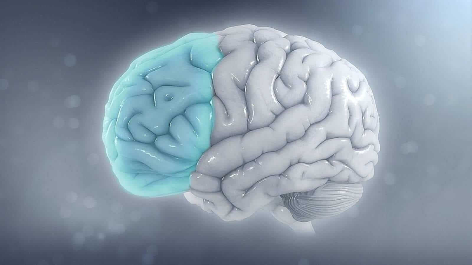
Under anesthesia, individual brain regions can still respond to the world. Researchers say what goes missing is the conversation among them.
Brain imaging studies show that anesthesia reduces overall brain activity, but researchers say the bigger change may involve communication. Connections between distant brain regions become less frequent, even though individual areas can keep responding.
In other words, the brain may not stop working. Instead, the coordinated exchange that allows thoughts, experiences, and awareness to emerge appears to weaken.
Andrea Luppi of the University of Cambridge has described consciousness this way: “During wakefulness, the stream of consciousness guides the sequence of brain patterns that we visit.” Under anesthesia, that guidance fades. Functional MRI research has also found that individual brains become less distinctive while sedated, appearing more similar to one another.
Why Researchers Are Taking This Into the Operating Room
The science of anesthesia has long outpaced the tools used to monitor it, and that gap is drawing serious attention from researchers who think patients deserve better.
Although anesthesia directly targets the brain, brain monitoring is not standard practice in most operating rooms. “Surprisingly, we’ve been doing anesthesia for more than 150 years now, but we only recently started to measure the brain,” Helfrich said to Futurity. “Measuring the brain, even today, is not the standard of care.”
Her team hopes improved monitoring could guide anesthesia toward brain states closer to natural sleep rather than deeper, coma-like patterns. Researchers are especially interested in whether better control could reduce cognitive and memory difficulties that sometimes affect older adults and patients with existing health challenges after surgery.
The Baylor team sees another possible path. The patterns they observed in the hippocampus may eventually help inform technologies such as speech prosthetics for people with certain brain injuries.
There is also a practical concern behind the research: accidental awareness during surgery. The event, in which a patient regains some level of consciousness during an operation, is estimated to occur in roughly one out of every 15,000 cases. It is uncommon, but with hundreds of millions of surgeries performed each year, even rare events affect many people.
Scientists still do not have a complete answer for what separates a functioning brain from a conscious mind during anesthesia. What is becoming clearer is that unconsciousness is not the same as inactivity. Beneath the silence, the brain can remain busy, responsive, and far more complicated than it appears.
Has learning that the brain stays more active during anesthesia than most people realize changed how you think about surgery, either for yourself or someone you love?